How is the nervous system organized?
The nervous system is one of the most complex and functioning systems in the human body. It unites and at the same time controls all processes in the human organism. Its in-depth study continues to this day, looking for various mechanisms that allow us to look deeper into human intelligence and capabilities.
The nervous system is divided into two parts – central and peripheral nervous system , which, despite their division, function as a whole. The central nervous system (CNS) is represented by the brain and spinal cord, while the peripheral – generally by nerves that reach all parts of the body. Nerves, like a network of receptors, provide information about various conditions and changes in the human body to the brain. This information is processed and causes a feedback, which is transmitted from the brain through the peripheral nerves to the relevant organs and systems.
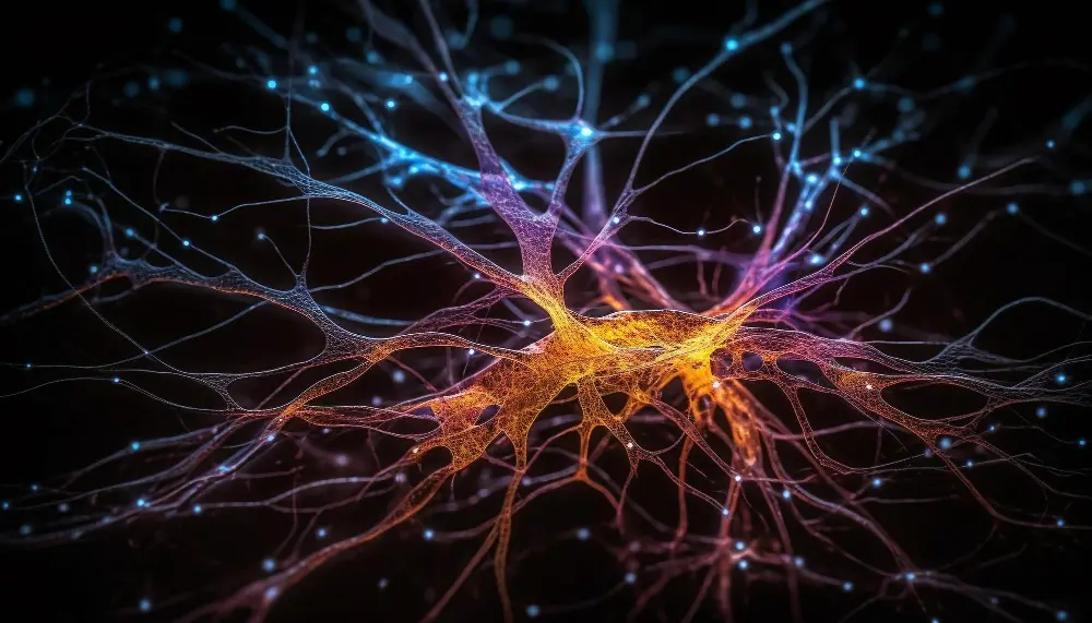
The main structural and functional unit of the nervous system is the nerve cell, or the so-called neuron . Neurons in the nervous system vary in shape and size, but the structure of all is similar. Their number is about 100 billion, and each neuron is connected to at least 10,000 others. A neuron has a body and processes . One of the processes is usually longer than the others and is called an axon . Through it, the cell sends information to the other cells surrounding it, and these cells can be both nervous and of another type, for example, muscle, glandular, etc. Some of the axons can be covered from the outside with a myelin sheath, which is interrupted in places by the so-called twitches of Ranvier.
In these pinches, the nerve impulse, carrying information, jumps and thus it is more quickly transferred to the neighboring cell. The speed with which the pulse moves along these growths is from 50-60 to 120 m/s. Other axons do not have such a sheath, and their speed is significantly slower – the lowest speed is on the order of 0.5 m/s. The remaining growths are shorter, they are called dendrites and with their help the given neuron receives information from other nerve cells. In general, the body of the neuron serves to process and integrate the information supplied.
The place of contact of two nerve cells is called a synapse (from Greek “son” – together). This is a narrow cleft between the axon of one neuron and the dendrite of another. A special chemical substance called a neurotransmitter or neurotransmitter (for short just a mediator) is released from the axon of one neuron, passes through the cleft (synapse) and reaches the dendrite of the other neuron. In this way, a signal and information are transferred between the two neurons.
There are many mediators in the nervous system – acetylcholine, dopamine, which serve to transmit various informational signals to neurons. Their diversity underlies the variety and complexity of nervous processes. A number of diseases are due precisely to a violation in their separation, synthesis or degradation. This shows that the nervous tissue is an extremely complex interwoven network in which cells are constantly interacting and exchanging information. Another type of cells take part in this network, whose function is more auxiliary than structural. This is the so-called glia – cells located between individual neurons that protect, nourish and support the functioning of neurons. Glial cells are significantly more diverse in shape and simpler in structure than neurons.
According to their function, neurons are:
- afferent (“incoming”, sensory), which transmit information from different parts of the body to the CNS,
- efferent (“output”, motor), which transmit the information from the central nervous system to the organs and systems of the body
- interneurons that make the connection between them.
What is the spinal cord?
The spinal cord is the connection between the brain and the peripheral nervous system . It is located in the spinal canal, reaching 2-3 lumbar vertebra. It looks like a string and is about 42-45 cm long. In cross-section, it has the shape of a butterfly, in which the gray matter of the brain, made up of the bodies of neurons, is located on the inside, while the white matter of the brain – represented by their axons, is on the outside. In the center is the spinal canal through which cerebrospinal fluid flows. 31 pairs of spinal nerves
emerge from the spinal cord , which belong to the peripheral nervous system by function. These nerves pass through special openings in the vertebrae and then reach the various tissues and organs. These include 7 cervical, 12 thoracic, 5 lumbar, 5 sacral and 1 coccygeal depending on the vertebra from which they arise. Each spinal nerve is formed by a posterior and anterior branch. The posterior branch branches off into various tissues and along it, “incoming” information from the relevant organs reaches the central nervous system.
The anterior branch is made up of the axons of neurons located in the gray matter of the spinal cord. Through it flows “outgoing” information from the central nervous system to the tissues and organs for the performance of various functions. The gray matter of the brain itself is composed of an anterior and a posterior horn. The posterior root (sensory, “incoming”) reaches the posterior horn, and the front root (motor, “outgoing”) begins from the anterior horn.
In general, the spinal cord is connected with the reflex activity of the body, the most familiar example of which is the knee reflex – when tapping under the knee cap, the information about this passed through the peripheral nerves and the sensory (incoming) root, it is also processed through the motor (outgoing) radicle and peripheral nerves reach the thigh muscles to cause extension in the knee.
How is the brain structured?
The brain is part of the CNS. It is located in the cranial cavity and thus is protected from blows and trauma in the head area. This protection is also helped by the fact that the brain is surrounded on all sides by liquid (cerebral cerebrospinal fluid) and is surrounded by meninges – membranes that additionally protect it from various shocks.
The brain is made up of gray and white brain matter . The gray matter is made up of the bodies of the neurons, while the white matter is made up of their axons, whose sparkling white color is characteristic. The weight of the brain is different, but most often in men it is about 1300-1350 g, while in women it is about 1250 g. Its volume is about 1,600 cm2. These differences are due to the greater amount of white brain matter in men compared to women. In composition, brain tissue is a fibrous, jelly-like mass that is very rich in water and lipids.
The brain is made up of the following parts:
- brain stem
- small brain
- Intermediate brain
- cerebrum
- ventricles
How is the brain stem structured?
It is the smallest and, from an evolutionary point of view, the oldest and most primitive structure of the brain. It is associated with vital functions such as breathing, feeding, maintaining heart rate and blood pressure, feeding. The brain stem is made up of several elements.
The medulla oblongata is located closest to the spinal cord. It houses vital centers such as the one for breathing, cardiac activity, and its damage is associated with irreversible and life-incompatible conditions.
The pons is the structure located slightly above the medulla oblongata. It plays the role of an intermediate station in the transfer of information between the cerebrum and the cerebellum.
The midbrain is the third element of the brainstem. It is important for head and eye movements, pupillary reactions, and skeletal muscle tension.
A special group of nerve cells is located in the brain stem, which are collectively called the reticular formation . They are of fundamental importance in maintaining the states of wakefulness and sleep, and in the event of disturbances in this system, pathological conditions such as insomnia can occur.
It is from the brainstem that almost all cranial nerves that belong to the peripheral nervous system originate. These are the oculomotor, trochlearis, trigeminus, abducens, facialis, vestibulocochlearis, glossopharyngeus, vagus, accessory and hypoglossus . Only the first and second cranial nerves – olfactory and optic – do not originate from the brainstem. They mainly control the functions of anatomical structures in the head area. The exceptions are the vagus, which supplies sensory information from the internal organs, and the accessory nerve, which innervates two muscles.
What is the cerebellum?
It occupies the posterior parts in the cranial cavity. It consists of two hemispheres, connected to each other , which have an uneven surface and are grooved. Its function is directly related to the coordination of body and eye movements, the control of muscle tone, the maintenance of a certain posture in space, and the provision of body balance.
This part of the brain is highly developed in birds, which is related to the fine tuning of muscles in flight.
What is the midbrain?
The midbrain is composed of the thalamus and hypothalamus .
The thalamus is the intermediate station of all the sensory pathways that come from the body and go to the brain. It processes the information that has reached it and sends a part of it to the brain, where a finer and more precise analysis can be carried out. Another piece of information is retained at this level.
The hypothalamus is a structure that weighs only 4 g, but is of vital importance to the entire organism. Located below the thalamus, it is the most important regulatory organ in the human body. It has both nervous and endocrine functions, both of which are aimed at ensuring the relative constancy of the body’s internal fluid environment. Near the hypothalamus is the main conductor of the endocrine glands, namely the pituitary gland. It is closely related to the hypothalamus.
How is the brain structured?
The brain is located at the top and makes up about 2/3 of the entire brain parenchyma. Its surface is striated and composed of numerous folds, which provides a greater number of cells in a significantly smaller area. If the folds of the brain could be smoothed out, their length would amount to half a square meter. The brain is composed of two hemispheres , which are connected to each other by a bundle of white brain matter. In this way, they are in constant contact.
An interesting fact is that the hemispheres control the opposite parts of the body , for example, the right hemisphere is responsible for the movements of the left half of the body, and vice versa. The outermost part of the brain is covered with the cerebral cortex – the highest structure that is characteristic only of humans. Its thickness is about 4 mm. It is related to the thought and analytical process and is the evolutionary highest step in the development of the nervous system.
The cortex of each hemisphere is composed of four lobes, each of which has a specific function. The frontal or frontal lobe is responsible for thinking and imagination, containing centers related to body movement as well as speech centers. Thanks to the parietal lobes, we recognize the objects around us, they also contain the sensory area, through which we perceive various sensory sensations. The temporal lobes are associated with hearing, understanding words, and the occipital lobes with vision.
Functionally, the CNS includes the so-called limbic nervous system . Its anatomical substrate is structures located in different parts of the brain, with the cerebral cortex playing an essential role. The limbic system is mainly related to emotions, pressure, instincts, memory. It participates in reactions related to the self-preservation of the individual and the species as a whole.
It is an interesting fact that although the brain makes up a very small part of the human body, it takes up about 20% of the blood flow in his body. It is the organ most sensitive to oxygen and glucose starvation. When the blood supply is interrupted for about 5 minutes, a person becomes unconscious, and after that time, irreversible changes in brain activity begin to occur.
The blood supply to the brain is carried out by two systems. One includes the two carotid arteries that run along the front of the neck. Each of them is divided into an external one, which supplies blood to the face, and an internal one, which nourishes 3/5 of the brain. In the brain itself, the internal divides into the anterior and middle cerebral arteries.
The second system is represented by the two vertebral arteries, which pass through special openings on the vertebrae. In the brain, they join to form the basilar artery, responsible for feeding the remaining 2/5 of the brain. It gives rise to the posterior cerebral artery and some branches that connect to the anterior and middle.
On the outside, the brain and spinal cord are covered by three membranes called meninges . These are the dura mater, the arachnoid and the soft meninges . The latter is in direct contact with the brain parenchyma. Cerebrospinal fluid flows between them, and they are essential for protecting the brain from various harmful factors.
What is the ventricular system?
The ventricular system is made up of 4 structures in the brain that are connected to the central canal of the spinal cord. They are called cerebral stomachs and are the left and right lateral, third and fourth cerebral stomachs . Cerebrospinal fluid is produced in them. From the two lateral stomachs, the cerebrospinal fluid enters the third cerebral stomach through the foramen of Monro.
From there, through the cerebral aqueduct, the cerebrospinal fluid enters the fourth cerebral stomach, which on the one hand is connected to the central canal of the spinal cord, and on the other, to the space between the arachnoid and the soft brain shell (the subarachnoid space). From the latter, the cerebrospinal fluid is reabsorbed using the venous system of the brain. When the path of movement of the liquor is disturbed and its retention in the stomachs, they expand and the picture of internal hydrocephalus is obtained.
What is the peripheral nervous system?
The peripheral nervous system connects the body with the central nervous system. Unlike the central nervous system, it is not protected by bones and thus is exposed to the harmful action of many factors. Information is continuously exchanged between the two systems via 31 pairs of spinal nerves (nerves connected to the spinal cord) and 12 cranial nerves (nerves connected to the brain). These nerves contain both motor fibers that control muscle function and sensory fibers that carry sensory information.
The anatomical division of the peripheral nervous system includes the afferent (incoming) and efferent (outgoing) part .
Functionally, however, the PNS is divided into somatic and autonomic.
The somatic nervous system is concerned with the conscious control of body movements, influencing skeletal muscle function. In addition, it also processes the sensory information from the various sense organs and the skin.
The autonomic nervous system is a system of nerves that conduct information in the form of nerve impulses from the walls of blood vessels, from the heart, from the internal organs, from the organs in the pelvis to the central nervous system and, in particular, to the medulla oblongata, to the pons and to the hypothalamus. These impulses remain unconscious, i.e. without the participation of consciousness and lead to the production of corresponding efferent impulses that affect the function of the specified structures and maintain their equilibrium. The autonomic system has two main components – the sympathetic and parasympathetic nervous systems .
The two systems have exactly the opposite effect on the body. The parasympathetic nervous system generally slows down the body’s functions, such as slowing the heart rate while a person is resting, lowering blood pressure, constricting the pupil. The sympathetic nervous system does the opposite – it turns on when a person is extremely active or under emotional stress. Under its influence, the heart rate and blood pressure increase, the pupil dilates, and sweating increases. The two systems function in a coordinated manner, without the involvement of consciousness. A peculiarity of their anatomy is that each of them consists of two neurons.
The first of them is located in the gray matter of the spinal cord or in the brainstem, while the other neuron is located outside the CNS and their number significantly exceeds that of the first neurons. In the sympathetic system, the two neurons are close to each other, while in the parasympathetic system, the second neuron is located next to or in the effector organs themselves. In addition, the two systems also differ in the localization of their first neuron, as well as in the mediators released by them. In the sympathetic nervous system, both neurons release acetylcholine, while in the parasympathetic neurons, only the first releases this mediator. The second works with the help of norepinephrine.


