Can we protect ourselves from arthrosis and how?
Since in the majority of cases, the cause of the occurrence of arthrosis is unclear, there are no sure methods and means of preventing its occurrence. However, several measures can be taken to reduce the occurrence of this disease.
All congenital and acquired conditions, which are known to cause arthrosis in a high percentage, should be detected in time, followed up, and treated. Some of them, for example, such as congenital deformities, can be cured definitively by surgical treatment.
Building such a lifestyle that reduces the risk of developing arthrosis – reducing body weight, avoiding extreme loads on the joints, and at the same time ensuring an optimal motor regime for good muscle strength and a good volume of movement, preventing osteoporosis through wholesome nutrition, optimal motor mode and, if necessary, hormone replacement therapy for women, etc.
What is the prognosis for recovery of joint function after treatment?
It is impossible to give a universal prognosis for the disease. It depends on the type of the affected joint, the number of affected joints, the cause of the arthrosis (if it is detected), the presence of risk factors, the timely and correctly conducted treatment, etc.
Usually, with well-conducted and timely treatment, arthrosis is rarely a debilitating disease. However, its course is chronic, prolonged, and with a constant tendency to worsen.
None of the means of treatment (except endoprosthetics to some extent) does not definitively cure this disease. As the disease progresses, it may be necessary to change both the treatment and the way of life (with the means of psycho-social rehabilitation).
What is the operative treatment?
When conservative treatment has exhausted its possibilities, operative (surgical) treatment is resorted to. It is also diverse. The most frequent operative interventions are the following:
arthroscopy of the joint with excision of part of the synovial membrane, filing of the spikes, smoothing of the cartilage, bosonization of the subcartilaginous bone to improve cartilage nutrition, etc.; it is usually a step toward more definitive operative treatment
synovectomy – a surgical removal of the chronically inflamed and altered joint capsule and synovial membrane, which to a large extent removes painful sensations from the joint and facilitates movements in it
arthrodesis – is “liquidation” of the joint by surgical means. The movable connection between the bones becomes immovable. The pain disappears completely, but the function of the joint is lost
chondroplasty – a modern operation in which one’s own or foreign cartilage is grafted to the place of the destroyed cartilage, which fills the defect. The results in this direction are encouraging. Genetically grown cartilage has recently been reported. Unfortunately, most techniques of this kind are considerably expensive
operations improving the mechanical function of the joint – these are corrective osteotomies for the limbs, cap plastics, etc.
endoprosthetics more popularly as “artificial joint ” – is an operation in which the extremely changed and destroyed joint is replaced with an artificial one made of various materials such as titanium, special steels, polyethylene, etc. Hip and knee joints are most often endoprosthetic, less often shoulder, elbow, ankle, and small joints of hands and feet.
What is conservative treatment?
Treatment options are numerous depending on the number and type of affected joints, the cause of the disease and its severity, the age and general condition of the patient, financial factors, etc. Treatment is usually staged. First, conservative treatment (anything other than surgical treatment) is started, and when it does not give results or when these are exhausted, surgical treatment is resorted to, if there are no contraindications for this.
If the cause of the arthrosis is known (secondary arthrosis), then the treatment is directed against it (if this is possible – e.g. operations for congenital deformities, incorrect mechanical axis of the limb, eradication of infection in the joint, etc.). If the cause is unclear (as it is in the majority of cases), the treatment aims to eliminate the risk factors (obesity, increased activity involving a given joint, insufficient nutrition, etc.) and relieve the symptoms (pain, swelling, limited movements) while preserving and improve joint function.
Usually, in mild cases, conservative treatment is started. In various combinations, its main elements are:
- lifestyle change – particularly important for arthrosis of large, weight-bearing joints is weight reduction. In many cases, this alone significantly relieves symptoms. It is important to avoid large, painful, and other complaints loads on the joint concerned. The rest of the joint should not be absolute, it is extremely important to maintain activity of the joint, but to a degree that does not cause pain.
- physiotherapy and rehabilitation – a specialist rehabilitator and physical therapist develop a program in which the main focus is on exercises to maintain a good range of motion in the joint and stabilization by strengthening the muscles. Various methods are used to relieve pain and swelling, such as cryotherapy (ice treatment) for newly activated arthrosis, via dynamic and interference currents, application of heat (warm showers, heating, paraffin treatment) for non-activated arthrosis, application of ultrasound, infrared rays, iontophoresis with medicines, etc.
- to ensure the rest of the joint, various orthotic devices can be used – splints, shoes with high and soft soles, corsets and collars (for the spine), crutches, canes, etc.
- anti-inflammatory drugs – usually they are non-steroidal anti-inflammatory drugs and can be administered in different ways (orally, topically as gels, intramuscular injections) Since one of their main side effects is damage to the gastric mucosa when treated orally, the new ones are preferred, selective representatives of this group of drugs that do not have this side effect. These drugs are sold without a prescription, but it is recommended to consult a doctor before starting to take them.
If the pain and swelling do not respond to this treatment, an intra-articular injection of a corticosteroid (a powerful anti-inflammatory agent) may be given, usually in combination with a local anesthetic. This method achieves pain relief for a fairly long period but carries certain risks.
- taking nutritional supplements containing the substances important for cartilage, chondroitin, and glucosamine sulfate. Their effect is not yet fully understood.
- taking drugs containing ingredients of normal cartilage, these are so-called chondroprotectors or substances protecting or strengthening articular cartilage. They mainly contain hyaluronic acid and some other glycosaminoglycans. These drugs can be taken orally (this is carried out according to a special scheme, in courses of different duration, and must be prescribed by a doctor) or administered directly inside the joint by introducing them with a syringe and needle. The injections are also given on a schedule, in courses, for varying periods, and usually achieve relatively long-lasting relief of symptoms (mostly pain).
The different methods of conservative treatment are usually applied in combinations for an optimal result.
How is the diagnosis made (how is the disease detected)?
The process of establishing the diagnosis begins from the first contact of the doctor with the patient. In a conversation with the patient, the doctor acquires information about the type and nature of his complaints. Answering his questions, you will report what kind of pain you feel, where it is the strongest, what makes it worse and what relieves it, how old the complaints are, whether have you had any congenital diseases of the bones, joints, or metabolism, have you suffered injuries to the musculoskeletal system, what is your profession, are you active in sports, how do you move, what medications have you taken so far and what was their effect, are there other members of your family who have the same complaints and so on..
The doctor will then examine you thoroughly, paying close attention to the joint or joints from which the complaints are most likely to originate. Compared with the healthy side (if only one of two symmetrical joints is affected), the doctor will look for signs of inflammation (swelling, redness, etc.), muscle weakness, shortening of one of his limbs, and a strange position of the same.
The doctor will examine the strength and volume of active (voluntary) and passive (performed by the doctor, without the participation of the patient’s muscles) movements in the affected joint. The doctor will analyze by looking at your posture and gait.
Usually, the information from the examination so far is sufficient as a guide to the most likely diagnosis. To confirm the diagnosis and to rule out some other possible diseases and conditions, the doctor will prescribe several instrumental tests.
When should you seek medical help?
The symptoms of arthrosis can be symptoms of other diseases, so it is recommended that your condition be clarified by a doctor.
Having joint or bone pain, loss of range of motion in a joint, joint swelling and warmth, fever and weight loss, reddening of the skin over the joint, shortening of the leg, and all of the above symptoms are signs that you need medical help.
What are the most common types of arthrosis?
The most common types of arthrosis (of the spine, of the hip joint, of the knee and of the fingers) are also characterized by some more specific symptoms for each of them:
- arthrosis of the spine
- arthrosis of the hip joint (coxarthrosis)
- arthrosis of the knee joint (gonarthrosis)
- arthrosis of the small joints of the hands
What are the main symptoms of arthrosis?
Arthritis affects only the joints and surrounding structures, this disease does not cause systemic complaints such as weight loss or increased temperature, nor does it cause direct complaints from the internal organs (unlike some of the arthritis ).
The symptoms of arthrosis may differ depending on which joint is affected, how many joints are affected, the age of the patient, the presence of accompanying diseases, etc. Each person has an individual pain tolerance.
The most common symptoms of arthrosis are the following:
- pain – the pain is usually dull and felt deep in the joint, it intensifies when performing physical activity involving the joint (at the beginning of the disease, pain is observed when the joint is overloaded, later during active movements and in the advanced stages – at rest ); pain can be provoked when the weather worsens; the pain may appear when moving the joint after a long rest (eg, knee pain when standing up after sitting for a long time); usually the pain subsides at rest, but in the more advanced stages of the disease it can become constant and so independent of physical efforts; the pain can also appear at night and sometimes wakes up patients from sleep; sometimes the pain, as well as some of the other complaints, can subside for various long periods (eg in the case of arthrosis of the small joints of the hands).
- stiffness in the joint – usually occurs after a long period of rest of the joint and disappears after movement, may progress over time
- limitation of movements in the joint – occurs gradually, sometimes it can prevail over the pain
- swelling of the joint – inflammation of the synovial membrane causes an effusion of fluid into the joint cavity.
- warming and possible redness of the skin over the joint – inflammation of the joint sheaths causes activation of all exchange processes inside the joint, which causes warming
- deformity of the joint and abnormal position of the limb or body – the contraction of the joint capsule and the surrounding muscles and tendons and the deformation of the bones causes conditions called contractures. In the presence of contracture, the limb takes an unnatural position or cannot perform the full volume of a given movement.
- hypotrophy (weakening) of the muscles performing movements in the affected joint; pain and limited mobility of the joint deprive the muscle of activity, which leads to its weakening.
- spasm of the surrounding musculature – occur in the early stages of the disease
- exposure of the subcartilaginous bone causes a sensation of “popping” and “crunching” in the joint during movement; the presence of loose “joint mice” in the joint cavity can cause the joint to become blocked
- specific symptoms in the various joints most often affected by arthrosis
Which joints are most often affected by arthritic changes?
Arthrosis of the spine ( spondylarthrosis ) is in first place in terms of frequency, followed by the involvement of the knee (in medicine it is called gonarthrosis ), hip (in medicine it is called coxarthrosis ), shoulder joint ( omarthrosis ), and the small joints of the hands. Arthrosis can affect one or more joints, and the involvement can be symmetrical.
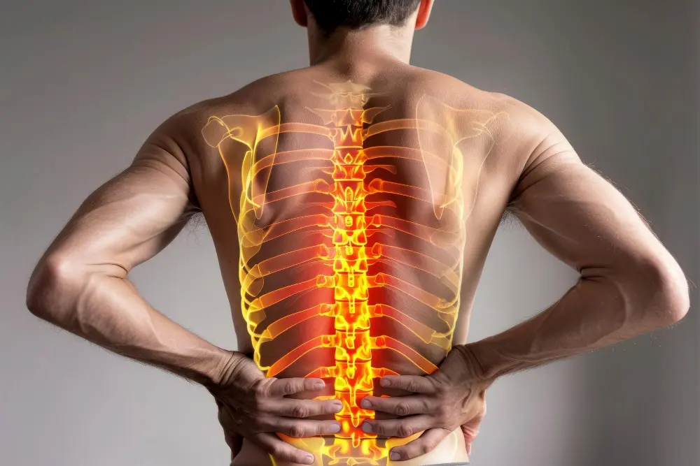
Affecting the spine is most common in the cervical and lumbar region, where the load is greatest.
The knee and hip joints are the ones that bear the weight of the entire human body. Often, the involvement is symmetrical (ie, both joints are affected).
Of the small joints of the hand, the joints between the closest to the wrist and the middle phalanx (the bones of the fingers are called phalanges) and the joints between the middle and the farthest phalanx from the wrist are most often affected. The characteristic of these cases is the formation of hard swellings around these joints, which are called nodes of Heberden and Bouchard. Heredity is present in this arthrosis and it occurs more often in women from one family.
Of the joints of the foot, the joint at the base of the big toe is most often affected, where a characteristic bony protrusion with a “callus” is formed on the outside.
With certain predisposing and causative factors, arthrosis can develop in any joint.
What changes in joint tissues does arthrosis cause?
Arthritic lesions of a joint are the result of a large number of disease processes, ultimately leading to a structural and functional failure (due to a final change in the structure, the joint cannot perform its function properly) of one or more joints. Osteoarthritis affects the entire joint, including the muscles and tendons located around the joint, the ligaments, the underlying articular cartilage, the synovial membrane that “upholsters” the joint, and the joint capsule that “envelops” the joint from the outside.
The primary lesion is in the articular cartilage. Under the action of the number of factors listed above, microcracks form in it and its elastic properties are disturbed. Gradually, the articular cartilage is destroyed, which leads to a narrowing of the joint gap (the cartilage serves as a “spring” between the contacting bones). In the final stages of cartilage destruction, the “bare” subcartilaginous bone underneath is exposed and friction occurs between the bony surfaces on both sides of the joint.
Tiny fractures occur in the subcartilaginous bone as a result of the altered load. From the crushed bone beams and pieces of destroyed cartilage, bodies are formed in the joint, which cause an outpouring of enzymes and inflammation of the joint “upholstery” – the synovial membrane. Sometimes these bodies can become rounded and remain freely mobile in the joint cavity and are called “joint mice”.
The inflammation of the synovial membrane causes an effusion of inflammatory fluid in the joint and the occurrence of edema, accompanied by pain and warmth in the joint area.
The changed mechanical load of the subcartilaginous bone (due to the absence or impaired function of the cartilage) leads to remodeling (the body tries to compensate for the unfavorable changes in the structure of the joint and preserve the function as long as possible). This reconstruction is taking place in several stages.
Initially, the body reacts with a new production of albeit altered bone. The sub-cartilaginous bone layer is compacted (sclerosis) and so-called bone spikes (osteophytes ) are formed on the edges of the joint surfaces, which represent outward protrusions. The spikes increase the area for transferring the load from one bone to the other and are a compensatory reaction of the body. In the more advanced stages of the disease, bone degradation changes prevail – cysts (rounded hollow formations in the bone filled with whitish fluid) appear in the bone tissue located immediately under the joint surface. Bone-on-bone friction reduces joint mobility and stability.
The inflammation of the synovial membrane leads to the growth and, subsequently, the inflamed joint capsule shrinks and limits the movements in the joint to varying degrees.
Due to the reduction of movements in a given joint, sometimes to the point of immobility (both due to mechanical obstacles and due to pain of varying intensity), contraction and shortening of the muscles located around the joint occur (they decrease in volume and decrease in length).
Contraction of the joint capsule and the surrounding muscles with their tendons leads to deformation of the joint and an abnormal position of the bones connecting in the joint. This condition is called contracture of the joint and is a sign of an advanced stage of the process.
Articular cartilage does not have nerve endings, i.e. its destruction does not cause pain in itself. The occurrence of inflammation of the synovial membrane and the joint capsule leads to the appearance of pain, swelling, and warmth. This is the moment when a “latent” or “silent” arthrosis becomes an “activated” arthrosis and begins to cause severe complaints. The cause of the pain is also the friction of the “bare” subcartilaginous bone, which is richly innervated.
As a rule, changes in the joint always exceed the degree of complaints expected in a given patient.
What causes osteoarthritis?
Despite the high frequency of this disease, not all the causes of its occurrence are sufficiently clarified. Primary or “idiopathic ” is called this arthrosis, the reasons for the development of which are not known. This is where the overwhelming percentage of cases fall. With this arthrosis, we can only talk about risk factors predisposing to the development of the disease but not directly causing it.
Primary arthrosis occurs mostly in elderly people (with advancing age, the water content of the articular cartilage increases, its protein components lose their qualities, microcracks gradually appear in the cartilage and its destruction occurs); it is known that women are affected 3 times more often than men by atrosis; it is assumed that diet (protein and salt composition of food) also has an influence; a greater frequency of arthrosis was observed in certain families, which suggests dependence on genetic factors causing congenital cartilage deficiency; the level of certain hormones at different times of life may also play an important role in the development of the condition; arthrosis of load-bearing joints develops more often in overweight people, etc.
Secondary arthrosis is referred to when the causes of its occurrence are known (that is, there is some other disease state, as a result of which arthrosis occurs in one or more joints). Some of the more important reasons for the development of arthrosis are:
- traumatic damage to the articular surfaces and bones – here can be counted both major traumas (fractures passing through the articular surface of a joint; traumas changing the mechanical load on the bones, operations on the bones in the joint area, frequent dislocations, damage to the supporting apparatus of the joint, etc.) as well as microtraumas (obtained mainly during frequent, intense loads on certain joints, which occurs very often in active sportsmen, etc.). Any trauma carries an increased risk of developing arthritis changes in a particular joint after a varying length of time.
- congenital diseases of the musculoskeletal system – this includes a large number of conditions such as dysplasia of the hip joints, congenital dislocation of the hip joint, different lengths of the legs at birth, congenital absence or deficiency of any of the bones, crooked legs, X- and O-shaped knees, etc. These conditions are a prerequisite for the early development of arthrosis.
- aseptic necrosis of bones – in this condition, due to several reasons, the blood supply to the bone is stopped, which it “dies”, it is crushed and this disrupts its mechanical properties and functions. The most striking example of such a condition is aseptic necrosis of the head of the femur, which leads to the development of arthrosis of the hip joint.
- inflammatory processes in the joint – this includes the various types of arthritis that can lead to the destruction of cartilage, such as infectious arthritis or chronic, severe, immune-related diseases such as rheumatoid arthritis and Bekhterev’s disease, which can cause arthritic changes in all joints.
- prolonged immobilization of a joint can also lead to the development of arthrosis
- deposition of crystals in the joints – the deposition of uric acid crystals in gout and calcium crystals in pseudogout leads to inflammation in the joint tissues and eventual destruction of the joint cartilage.
- metabolic diseases (diseases of metabolism) – examples of such diseases that can be the cause of arthrosis are Marfan’s syndrome, Ehlers-Danlos syndrome, Wilson’s disease, hemochromatosis, Paget’s disease affecting bones, etc..
- frequent effusions of blood in the joint – this leads to an inflammatory process in the joint, causing further arthrosis changes; such bleeding is present in hemophilia and other blood clotting disorders.
- endocrine diseases (diseases of the endocrine glands that produce the hormones) – hyperparathyroidism, hypothyroidism (thyroid diseases associated with more and less than normal gland functions), diseases of the pituitary gland (produces growth hormone) and diabetes are conditions predisposing to the development of arthrosis. It is suggested that joint damage in diabetes occurs due to impaired “information reception” from the joint due to nerve damage from “diabetes”.
- osteoporosis is also an important risk factor
Most likely, the causes of arthrosis are combined and act in a complex manner (some as predisposing factors and others as more obvious causes).
What is osteoarthritis?
Arthrosis is a disease of one or more joints and occurs when the articular cartilage is damaged or worn. After the articular cartilage is affected, there is secondary damage to the underlying bone and inflammation of the joint shell (capsule), accompanied in most cases by the contraction.
The German-speaking authors use the term ” arthrosis ” and emphasize the degenerative (wear-related) changes in the joint structures and mainly the cartilage, while the Anglo-Saxon authors use the term ” osteoarthritis ” for the same condition, emphasizing the clinical manifestation of the disease, corresponding to inflammatory changes in the synovial covering of the joint, accompanying at certain stages the wear of the cartilage.
It is the most common arthritis, affecting millions of people worldwide. Before the age of 45, more men are affected, while after the age of 55 the female gender predominates.
Every person develops arthritic changes and these can be observed during an X-ray examination in 50% of people over 40 years of age, and in practice, there is no person over the age of 65 without arthritic changes in one or some of the joints. However, only 25% of affected people have complaints.
Прочети още на: Артроза | Puls.bg – https://www.puls.bg/illnes/issue_151/part_12.html


